This year’s American Association of Physicists in Medicine (AAPM) annual conference features a robust lineup of students and faculty from the Department of Medical Physics.
Alexandra Christensen, Laura Castañeda-Martínez and Lizbeth Ayala-Dominquez, PhD, are three members of the Quantitative Ultrasound Lab who will be presenting this year. Their work includes a phantom designed to simulate collagen in the cervix, new calibration methods for transducers, and non-invasive methods to detect liver fat in children.
A program highlighting the university’s participation is also available online.
Alexandra Christensen
Sunday, 1:58 p.m.| Room 209
An Adjustable Wool Fiber Phantom for Investigating the Detection of Anisotropy with Ultrasound Speckle Statistics
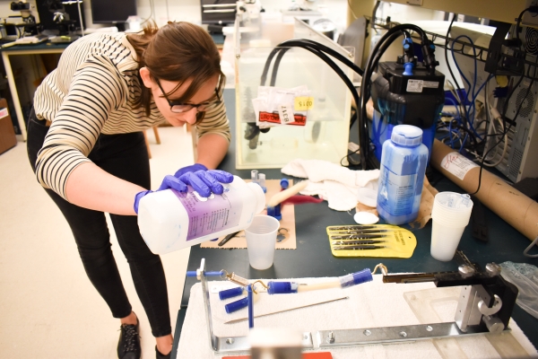 One of the major projects in the Quantitative Ultrasound Lab is the application of quantitative ultrasound to monitor the remodeling process in the cervix that precedes delivery to better understand and predict preterm birth. This AAPM presentation will showcase a phantom that was created using wool to simulate collagen in the cervix.
The problem addressed in this presentation is that models for the biomarkers used assume spherical media, not fibers like collagen. The goal is to better understand how these biomarkers change in fibrous media and work out new methods of interpretation.
The phantom was created using merino wool as the main material. Although not conventional for ultrasound phantoms, it has several properties (e.g. size, density, elasticity) similar to collagen. We can also change how aligned bundles of wool fibers are by stretching them out with a spring. We showed that if we image the phantoms from multiple angles, the biomarkers are able to distinguish not only between fibrous versus spherical material, but between stretched and relaxed fibers.
The presentation at AAPM will focus on the phantoms primarily, but there are more experimental details and preliminary in vivo results are available in an accepted manuscript.
Laura Castañeda-Martínez
Tuesday, 7:30 a.m. | Room 206
Best in Physics Ultrasound: A Calibration Framework for Quantifying Element Misalignment in 2D Matrix Array Transducers Used in 3D Quantitative Ultrasound
The goal of this project is to ensure accurate 3D ultrasound imaging by addressing a subtle but impactful challenge: micrometric misalignment in the elements of 2D matrix array transducers. It introduces a phantom-based calibration method designed to detect and compensate for small deviations in 2D matrix array transducers, improving both image reconstruction and measurement reliability. These novel transducers contain over a thousand individual elements, and even small shifts in their positioning can affect image quality and the accuracy of quantitative measurements. The source of this misalignment is linked to the manufacturing process, built into production and is difficult to correct before clinical or research use.
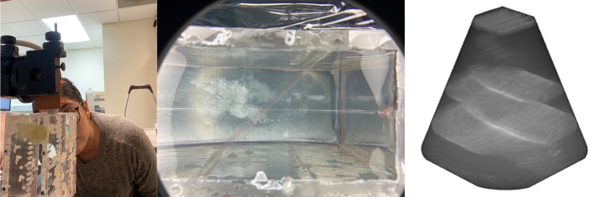
The phantom shown below was designed by Cristel Baiu. It is a multipurpose tool that includes over 50 nylon and tungsten fibers arranged at various depths and angles. It can be used to assess spatial resolution, slice thickness, and element misalignment. The tungsten section was used for the 2D matrix array calibration, which has a cross-hair target and three parallel fibers spaced 1 cm apart. This design enables reproducible and precise evaluation of element alignment, as well as other imaging quality metrics.
Lizbeth Ayala-Dominguez, PhD
Tuesday, 8 a.m. | Room 206
Preliminary Evaluation of Ultrasound-Derived Fat Fraction Reliability in a Pediatric Cohort
 Metabolic dysfunction-associated steatotic liver disease (MASLD) is the leading cause of chronic liver disease in children. While biopsy remains the diagnostic standard, non-invasive imaging parameters such as magnetic resonance imaging-proton density fat fraction (MRI-PDFF) and ultrasound-derived fat fraction (UDFF) are emerging methods for MASLD assessment. However, their clinical application in children is limited by challenges such as motion artifacts and a lack of standardized protocols. This study evaluates the inter- and intra-operator variability of UDFF in a pediatric cohort and its agreement with MRI-PDFF.
The goal of this project is to develop non-invasive ways to detect liver fat in children using ultrasound, as a more accessible alternative to biopsies or MRI scans that require breath-holding, which can be difficult for kids. “Preliminary Evaluation of Ultrasound-Derived Fat Fraction Reliability in a Pediatric Cohort,” focused on the ultrasound-derived fat fraction (UDFF), a measurement that uses a special algorithm available on Siemens ultrasound scanners. It found that UDFF measurements were consistent across different operators and when repeated on the same patient.
It found the measurements were more variable in children with higher body mass index, which suggests we may be able to refine the method even further. Next steps are to look at ways to adjust for differences in tissue properties and reduce that variability, hopefully making the technique even more accurate and useful for diagnosing liver disease in children.