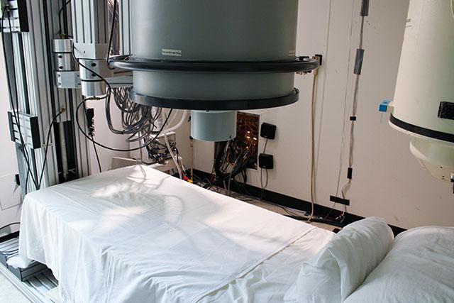
Biomagnetism
This is an accordion element with a series of buttons that open and close related content panels.
Ron Wakai, PhD | Biomagnetism Lab
Dr. Wakai conducts research in fetal MCG and in fetal, neonatal, and adult MEG. Research training is one of the primary missions of the lab.
Biomagnetism Lab
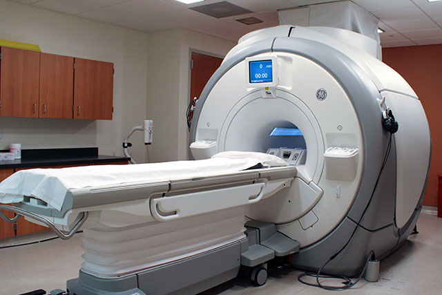
Magnetic Resonance
This is an accordion element with a series of buttons that open and close related content panels.
Andrew Alexander, PhD | Waisman Brain Imaging
Dr. Alexander’s research is focused on the use of advanced Magnetic Resonance Imaging (MRI) for mapping and measuring brain structure, tissue microstructure and functional organization. These techniques are being used to investigate the brain in both typically developing individuals and subjects across the lifespan.
Quantitative Brain Imaging Technology Lab
Walter Block, PhD
Dr. Block’s research interests include MR angiography and cardiac imaging – specifically designing real-time magnetic resonance acquisition and processing algorithms and systems for these procedures.
Dr. Block Profile
Doug Dean, PhD | Developing Brain Imaging Lab
Dr. Dean’s research focuses on the development and application of novel quantitative magnetic resonance imaging methods to measure and evaluate the brain structure throughout early development and aging. Current research is focused on examining how the white matter microstructure of the brain develops across early development and how these microstructural processes are related to changes in cognition and brain development in neurodevelopmental disorders.
Developing Brain Imaging Lab
Carri Glide-Hurst, PhD
Dr. Glide-Hurst’s primary areas of research and clinical expertise include magnetic resonance simulation (MR-SIM) and MR-guided radiation therapy.
Diego Hernando, PhD | Quantitative Imaging Methods Lab
By measuring physically meaningful properties of tissue, Dr. Hernando’s group aims to develop quantitative imaging biomarkers to improve the detection, staging and treatment monitoring of various diseases.
Kevin Johnson, PhD
Dr. Johnson’s work is focused on enabling the potential of MRI by accelerating acquisition speed, removing ambiguities and artifacts, and providing novel techniques for disease quantification. Through these advances, Johnson aims to help develop a new era of quantitative and molecular imaging across a broad spectrum of diseases. Some research interests include:
- MR pulse sequence development, non-Cartesian and non-Fourier imaging
- Signal encoding and decoding, sampling theory, recovery from incomplete samples
- Motion robust imaging, free breathing MRI, motion sensing
- Macro and micro vascular remodeling, perfusion, flow
- Improving the MRI experience for patients
Alan McMillan, PhD | Molecular Imaging/Magnetic Resonance Technology Lab
Dr. McMillan’s group seeks to advance MRI, PET/MR, and PET/CT imaging techniques and technology to gain new structural and functional information to better detect and assess human disease.
Beth Meyerand, PhD | Applied Neuro MRI Lab
Dr. Meyerand’s research is in the field of magnetic resonance imaging (MRI) of the human brain. The goal is the development of new MRI methods to visualize the structure and function of the brain and to translate these methods to the hospital for clinical diagnosis.
Marty Pagel, PhD | Contrast Agent Molecular Engineering Lab
The Contrast Agent Molecular Engineering Lab (CAMEL) directed by Dr. Pagel is developing MR Fingerprinting for small animal imaging studies. The CAMEL Group has developed Dynamic Contrast Enhanced (DCE) MRF that improves the quantitative imaging of vascular perfusion, and contrast agents for MRF that measure tumor acidosis.
Jim Pipe, PhD | Magnetic Resonance Technology and Use Design
The Magnetic Resonance Technology and Use Design (MRTUD) group focuses on building technology that is broadly impactful and holistically designed. From our core expertise in signal processing, math, and physics, we develop novel ways to sample data and reconstruct MRI images that are optimized for clinical impact. We design this technology in an open-minded, user-centric fashion, and combine it with approaches to greatly improve the operation and utilization of MRI in clinical settings.
Oliver Wieben, PhD | Cardiovascular MRI
- Cardiovascular MRI and MR Angiography: methodolgy development and translational research in data acquisition, image reconstruction, and post-processing
- Velocity-sensitive MRI: 2D and 4D Flow MRI to facilitate non-invasive hemodynamic assessment of the vascular system and CSF flow
- Rapid MRI: sequence design, reconstruction algorithms, and real-time imaging
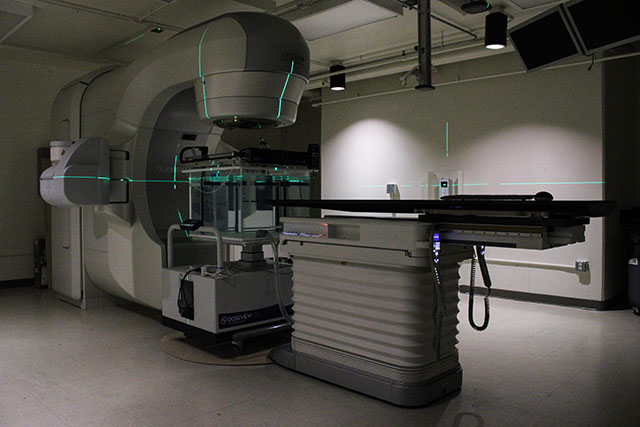
Metrology
This is an accordion element with a series of buttons that open and close related content panels.
Wesley Culberson, PhD | Wisconsin Medical Radiation Research Center
Research Interests
- Metrology
- Ionization chambers
- Radiation therapy
- Radiation dosimetry in diagnostic radiology and radiotherapy
- Thermoluminescent Dosimeters (TLDs)
- Calorimetry
Larry DeWerd, PhD | Wisconsin Medical Radiation Research Center
Radiation metrology, including calibration of ionization chambers, calorimetry, free air chambers. Radiation dosimetry in diagnostic radiology brachytherapy, and radiotherapy, quality assurance in radiology and radiotherapy, luminescence for dosimetry (TLD).
Wisconsin Medical Radiation Research Center
Ahtesham Khan, PhD | Wisconsin Medical Radiation Research Center
The focus of Khan’s current research is FLASH radiotherapy, which is a recently rediscovered mysterious effect that appears to reduce radiation-induced side effects when radiation is delivered in less than a fraction of a second. The lab is focusing on creating better detection methods for such radiation beams and performing pre-clinical studies to investigate the mechanism behind the FLASH effect.
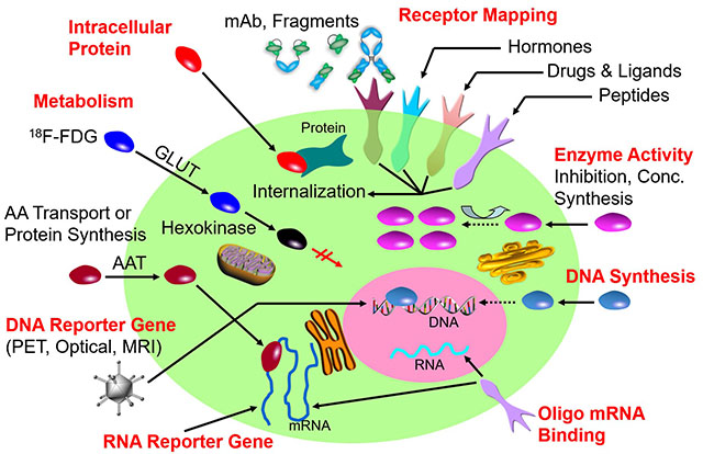
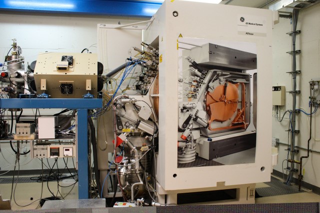
Molecular Imaging
This is an accordion element with a series of buttons that open and close related content panels.
Weibo Cai, PhD | Cai Research Group
Research of the Cai Group is focused on three areas: 1) development of multimodality molecular imaging agents; 2) molecular therapy of cancer; and 3) nanotechnology and its biomedical applications. Although our major disease focus is on cancer, the key feature of molecular imaging/therapy is that it is molecular specific instead of disease specific. The same molecularly-targeted agents can be applied in various diseases as long as the particular target is present or over-expressed. In addition, we are collaborating with a number of research groups both within UW and at other universities, where imaging can serve as an invaluable tool to answer the specific research questions posed.
Brad Christian, PhD | Brain Imaging
Dr. Christian’s research focuses on developing and translating novel PET methods for the study of neurodevelopment and neuropsychiatric illness. This involves using PET methodologies to investigate neurochemical changes in the brain and studying novel radioligands to characterize neurotransmitter-protein interactions and how they are influenced by development, genes, environment and drugs. These imaging methods are being applied to investigate the etiologies and mechanisms in diseases such as Down syndrome, affective disorders, schizophrenia, Alzheimer’s disease and Tourette syndrome.
Paul Ellison, PhD | Nuclear and Radiochemistry Lab
Dr. Ellison’s research interests utilize the principles and techniques of nuclear and radiochemistry to address challenges in the field of nuclear medicine. Drawing from the fields of nuclear chemical elemental separations, small molecule synthetic radiochemistry, preclinical cancer models, and preparation of radiopharmaceuticals for human research, Ellison’s work focuses on the development of small molecules radiolabeled with matched positron-emitting and Auger-electron-emitting therapeutic radionuclides for the planning and execution of targeted radionuclide therapy of cancer.
Reinier Hernandez, PhD | Advanced Radiotheranostics Laboratory
Dr. Hernandez’s research focuses on developing novel radiopharmaceuticals for therapy and diagnostics of cancer and other diseases. His research group emphasizes benchtop-to-bedside research to translate fundamental discoveries into life-saving technologies.
Advanced Radiotheranostics Laboratory
Robert Jeraj, PhD | Image Guided Therapy Group
Dr. Jeraj’s research focus is to improve cancer treatment by furthering the understanding of treatment resistance and the impact of tumor heterogeneity on treatment response using molecular imaging. We analyze patient data obtained from clinical trials to implement imaging biomarkers describing heterogeneity to understand both the development of treatment resistance and patient survival. Using the clinical trial data we aim to create models, machine-learning or computational, that further the understanding of disease progression.
Alan McMillan, PhD | Molecular Imaging/Magnetic Resonance Technology Lab
Dr. McMillan’s group seeks to advance MRI, PET/MR, and PET/CT imaging techniques and technology to gain new structural and functional information to better detect and assess human disease.
Marty Pagel, PhD | Contrast Agent Molecular Engineering Lab
The Contrast Agent Molecular Engineering Lab (CAMEL) directed by Dr. Pagel is developing molecular imaging methods that interrogate the tumor microenvironment, including tumor acidosis, hypoxia, vascular perfusion and enzyme activity, using MRI, PET/MRI, and photoacoustic imaging. In addition, the CAMEL group is developing contrast agents for intraoperative imaging, thermoablation and theranostics.
Nuclear Medicine
This is an accordion element with a series of buttons that open and close related content panels.
Brad Christian, PhD | Brain Imaging
Dr. Christian’s research focuses on developing and translating novel PET methods for the study of neurodevelopment and neuropsychiatric illness. This involves using PET methodologies to investigate neurochemical changes in the brain and studying novel radioligands to characterize neurotransmitter-protein interactions and how they are influenced by development, genes, environment and drugs. These imaging methods are being applied to investigate the etiologies and mechanisms in diseases such as Down syndrome, affective disorders, schizophrenia, Alzheimer’s disease and Tourette syndrome.
Paul Ellison, PhD | Nuclear and Radiochemistry Lab
Dr. Ellison’s research interests utilize the principles and techniques of nuclear and radiochemistry to address challenges in the field of nuclear medicine. Drawing from the fields of nuclear chemical elemental separations, small molecule synthetic radiochemistry, preclinical cancer models, and preparation of radiopharmaceuticals for human research, Ellison’s work focuses on the development of small molecules radiolabeled with matched positron-emitting and Auger-electron-emitting therapeutic radionuclides for the planning and execution of targeted radionuclide therapy of cancer.
Marina Emborg, MD, PhD | Preclinical Parkinson's Research Center
Dr. Emborg’s research focuses on understanding and developing treatments for neurological disorders, in particular Parkinson’s disease, in a highly collaborative, interdisciplinary fashion. Their work applies advance imaging techniques to minimize invasive procedures, maximize data collection and create new clinical applications as they search for safe neuroprotective strategies that will prevent, slow down or stop death of brain cells, and use stem cells as model systems and as cell–based strategies. The goal is to find solutions for neurodegenerative disorders and bring them to the clinic.
Jon Engle, PhD | Cyclotron Research Group
Dr. Engle’s work is actively pursuing novel accelerator targetry and radiochemistry for the production of radionuclides that are useful in medical diagnosis, disease treatment, basic science and industrial applications.
Reinier Hernandez, PhD | Advanced Radiotheranostic Laboratory (ART)
Advanced Radiotheranostic Laboratory (ART)
- Radiochemistry applications in biomedical research
- Targeted radionuclide therapy – theranostics
- Radio-immunooncology
Neutron capture therapy
Robert Jeraj, PhD | Image-Guided Therapy Group
Dr. Jeraj’s main research focus is to improve cancer treatment by furthering the understanding of treatment resistance and the impact of tumor heterogeneity on treatment response using molecular imaging. Their work analyzes patient data obtained from clinical trials to implement imaging biomarkers describing heterogeneity to understand both the development of treatment resistance and patient survival. Using the clinical trial data they aim to create models, machine-learning or computational, that further the understanding of disease progression.
Optical
This is an accordion element with a series of buttons that open and close related content panels.
Paul Campagnola, PhD | Campagnola Laboratory
- Biophotonics
- Tissue imaging
- Breast and Ovarian Cancer
- Connective Tissue Disorders
- Nanofabrication
- Cancer Cell Biology
- Tissue Engineering
Kevin Eliceiri, PhD | Laboratory for Optical and Computational Instrumentation
The Eliceiri lab develops new biomedical imaging and analysis methods to understand the roles of cells in disease. Projects include developing measures that can characterize the progression of cancer, building instruments to collect richer data than traditional methods, using machine learning to classify biomedical images, and developing open-source software for use by the image analysis community around the world. We are co-leaders in several major software development and education initiatives including the NIH P41 funded Center for Open Center for Open Bioimage Analysis (COBA), ImageJ/FIJI, Micro-Manager and the Scientific Community Image Forum. We are partners in several collaborative consortiums including the Bio-Formats project which originated in our lab and is now part of the Open Microscopy Environment (OME).
Melissa Skala, PhD | Skala Lab
The Skala lab develops biomedical optical imaging technologies for cancer research, cell therapy, and immunology. Current projects focus on tumor immunology and immunotherapy, cell-level metabolic heterogeneity, and cell-cell interactions. Collaborative projects leverage unique photonics-based tools for clinical problems, including quality control in T cell and stem cell therapies, designing personalized treatment plans for cancer patients, monitoring diseases in the eye, discovering new therapies for a range of diseases, and many others. Projects are highly diverse and range from translational research to hypothesis-driven questions to algorithm / instrumentation development.
Skala Lab
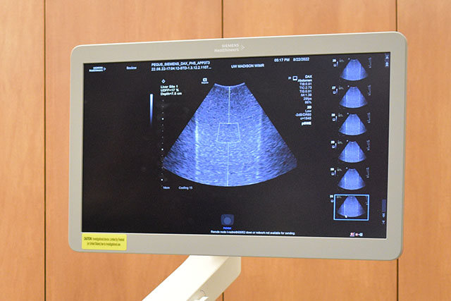
Ultrasound
This is an accordion element with a series of buttons that open and close related content panels.
Ivan Rosado-Mendez, PhD | Quantitative Ultrasound Lab
Dr. Rosado-Mendez’s research focuses on using ultrasound imaging to bridge the gap between microscopic and macroscopic imaging and understand how tissue properties at various spatial scales are related to each other and define the way biological tissue works and responds to a disease or lesion. By combining several sources of quantitative multiscale information, his ultimate goal is to develop ultrasound-based “fingerprints” describing the morphological, functional, and molecular properties of tissue. These fingerprints could help in the personalization of patient management, resulting in better outcomes. Furthermore, because of its safety, practicality, and portability ultrasound could help extend the benefit of personalized medicine to more patients.
Quantitative Ultrasound Lab
Tomy Varghese, PhD | Ultrasound Elastography and Photoacoustic Imaging Laboratory
Dr. Varghese’s laboratory research focus is the development of novel elasticity imaging techniques, quantitative ultrasound and clinical applications based on these approaches. They have developed elasticity imaging techniques for monitoring minimally invasive ablative therapies, shear wave imaging, normal and shear strain estimation with beam steered data, vulnerable plaque characterization, arterial stiffness measurements, cardiac elastography, uterine and cervical strain imaging and parametric imaging of quantitative ultrasound features.
Ultrasound Elastography and Photoacoustic Imaging Laboratory
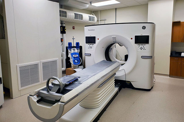
X-Ray/CT
This is an accordion element with a series of buttons that open and close related content panels.
Guang-Hong Chen, PhD | Dr. Chen's Lab
The application of cutting edge techniques and knowledge from the fields of physics, mathematics and computer science to medical disciplines has and will continue to generate innovations for the diagnosis and treatment of human diseases.
The mission of Dr. Chen’s laboratory is to apply x-ray physics, innovative image reconstruction methods and advanced computer technology to develop novel diagnostic tools for radiologists.
Frank Ranallo, PhD
Dr. Ranallo’s research involves the improvement and optimization of x-ray diagnostic imaging.
Research Interests:
- Dual energy CT
- Assistance in the optimization of hardware and software used in Radiographic, Fluoroscopy, Angiographic, and CT imaging systems.
- Creation of optimized CT scan and reconstruction protocols for multiple models of CT scanners: optimizing image quality at a given dose for the complete range of clinical indications.
- Optimizing protocols programmed into Radiographic, Fluoroscopic, and Angiographic systems.
Michael Speidel, PhD | Image Guided Interventions Lab
The mission of Dr. Speidel’s laboratory is to develop novel imaging tools for interventional radiologists and cardiologists through the application of physics, advanced imaging technology, and innovative algorithms.
Tim Szczykutowicz, PhD
Research Interests
- Optimizing CT scan protocols
- Monitoring patient dose
- Developing new metrics to define image quality in the clinical setting
- Developing protocol management methodologies
- Fluence field modulated CT
- Dual energy CT
- Cone beam CT