
Published on January 28, Enhanced porphyrin-based hypoxia imaging by temporal oversampling of delayed fluorescence signal, is a systematic study on how to enhance real-time imaging of tissue hypoxia through PpIX. Marien Ochoa, PhD, first author on the study and a scientist in the MOXI laboratory under Professor Brian Pogue, provided a brief overview of the study and goals for future research.
Real-time imaging of hypoxia dynamics in vivo has been a challenge, but Dr. Pogue’s lab has pioneered the use of endogenous PpIX for this purpose.
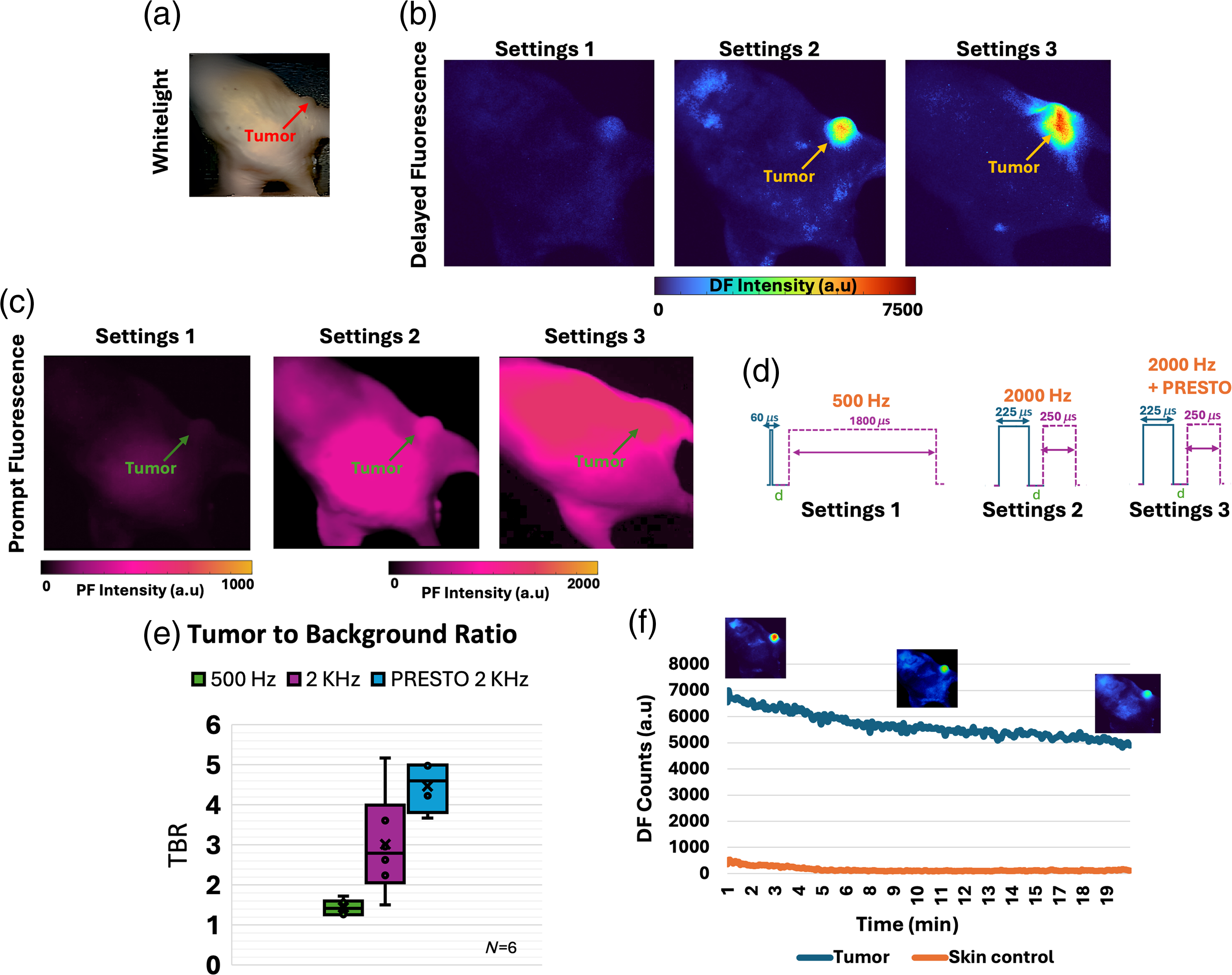
“This study explored enhancing PpIX DF signal intensity, which indicates tissue hypoxia, for real-time imaging of metabolism and hypoxia events. By increasing sampling frequency with a shorter gate width and longer pulse width, signal intensity improved approximately sevenfold in vitro and in vivo. This enhancement allows for better visualization of low-intensity hypoxic structures.
Furthermore the target-to-background contrast improved and is compatible with the previously reported PRESTO technique which further strengthens the magnitude of the detected signal. The study also examined photobleaching effects, showing that signal output remained strong even after prolonged exposure. Future research aims to image fast anatomical oxygen transients in real-time. The technique is currently being used to enhance contrast and understand hypoxia dynamics in tissues like tumors and lymphatic structures.”
Click for full image.