- Assessing Cervical Remodeling During Pregnancy
- Quantifying Backscatter Anisotropy
- Differentiating Types of Breast Masses
- Monitoring the Effects of Anesthesia on Neonates
- Assessing Renal Health
Summary:
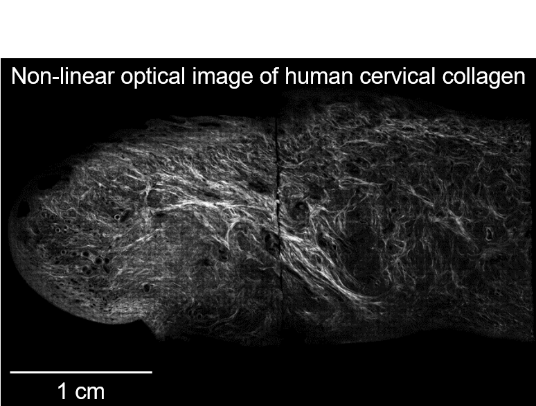
The cervix, or the lower portion of the uterus that extends into the vagina, acts as a final gatekeeper in pregnancy. A major constituent of this tissue is collagen and has been demonstrated to change extensively during gestation in animal models. Through use of our Quantitative Ultrasound approaches, we are investigating the normal biomechanical and microstructural changes that occur in the cervix throughout pregnancy.
Representative publications:
- Guerrero QW et al., “Anisotropy and Spatial Heterogeneity in Quantitative Ultrasound Parameters: Relevance to the Study of the Human Cervix,” Ultrasound Med Biol 44(7): 1493 – 1503. 2018.
- Carlson LC et al., “Detection of Changes in Cervical Softness Using Shear Wave Speed in Early versus Late Pregnancy: An In Vivo Cross-Sectional Study,” Ultrasound Med Biol 44(3): 515 – 521, 2018.
- Rosado-Mendez IM et al., “Quantitative Assessment of Cervical Softening During Pregnancy in the Rhesus macaque with Shear Wave Elasticity Imaging,” Phys Med Biol 63: 085016 (12pp), 2018.

Summary:
A common assumption in estimating various Quantitative Ultrasound parameters is that samples of interest are composed of isotropic scatterers. In reality, many tissues exhibit angle-dependent, or anisotropic, acoustic properties. Our group has been developing tools to quantify the magnitude of anisotropy and extract microstructural features. Initial tests of these parameters in the biceps muscle, a known anisotropic tissue, suggest these parameters are sensitive to differences microstructural organization and may be useful for for quantifying “normal” tissue architecture.
Representative publications:
- Guerrero QW et al., “Quantitative Ultrasound Biomarkers Based on Backscattered Acoustic Power: Potential for Quantifying Remodeling of the Human Cervix During Pregnancy,” Ultrasound Med Biol 45(2): 429 – 439, 2019.
- Guerrero QW et al., “Anisotropy and Spatial Heterogeneity in Quantitative Ultrasound Parameters: Relevance to the Study of the Human Cervix,” Ultrasound Med Biol 44(7): 1493 – 1503, 2018.

Summary:
While ultrasound can improve the sensitivity in breast cancer diagnosis over tests like mammography alone, improvements in its specificity are necessary for differentiating between benign and malignant breast masses. Our group has been investigating the use of elastography as well as backscatter-based Quantitative Ultrasound parameters to accomplish this.
Representative publications:
- Nasief HG et al., “A Quantitative Ultrasound-Based Multi-Parameter Classifier for Breast Masses,” Ultrasound Med Biol, In Press.
- Wang Y et al., “Three-dimensional Ultrasound Elasticity Imaging on an Automated Breast Volume Scanning System,” Ultrason Imaging 39(6): 369-392, 2017.
- Liu T et al., “Noninvasive In-Vivo Quantification of Mechanical Heterogeneity of Invasive Breast Carcinomas,” PLoS One 10(7): e0130258, 2015.

Summary:
Various studies have demonstrated that Quantitative Ultrasound is sensitive microstructural changes resulting from apoptosis. Through collaborations with physicians in neurology, we are investigating the ability of parameters like effective scatterer size (ESS) to correlate with apoptosis in the neonatal brain resulting from use of anesthesia.
Representative publications:
- Rosado-Mendez IM et al., “Quantitative Ultrasound and Apoptotic Death in the Neonatal Primate Brain,” Neurobiol Dis 127: 554-562, 2019.

Summary:
Kidney disease affects millions of people in the United States alone and represents a huge economic burden. Historically, clinicians would compare the speckle texture between the liver and kidney (see images above) to qualitatively assess renal health. Our group applied Quantitative Ultrasound parameters to quantify changes in renal microvasculature to objectively describe disease processes.
Representative publications:
- Hall TJ et al., “Ultrasonic Measurement of Glomerular Diameters in Normal Adult Humans,” Ultrasound Med Biol 22(8): 987-997, 1996.
- Wear KA et al., “Measurements of Ultrasonic Backscatter Coefficients in Human Liver and Kidney In Vivo,” J Acoust Soc Am 98(4): 1852-1857, 1995.
- Insana MF et al., “Identifying acoustic scattering sources in normal renal parenchyma from the anisotropy in acoustic properties,” Ultrasound Med Biol 17(6): 613-626, 1991.