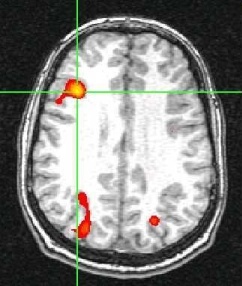
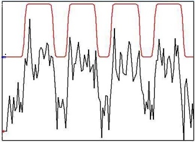
FMRI is a method of measuring the flow of oxygenated blood in the brain (Ogawa et al., 1990A, 1990B; Bandettini, 1992). FMRI is based on the BOLD effect where BOLD stands for blood oxygen-level dependent. BOLD MRI is accomplished by first exposing a patient or volunteer to a stimulus or having them engage in a cognitive activity while acquiring single-shot images of their brain. The region of the brain that is responding to the stimulus or is engaged in the activity will experience an increase in metabolism. This metabolic increase will require additional oxygen. Therefore, there will be an increase in oxygenated blood flow (oxyhemoglobin) to the local brain area that is active. Oxyhemoglobin differs in it’s magnetic properties from deoxyhemoglobin. Oxyhemoglobin is diamagnetic like water and cellular tissue. Deoxyhemoglobin is more paramagnetic than tissue so it produces a stronger MR interaction. We take advantage of these differences between oxy and deoxyhemoglobin in BOLD imaging by acquiring images during an “active” state (more oxyhemoglobin) and in a “resting” state (more deoxyhemoglobin). We see a signal increase in the “active” state and a signal decrease in the resting state. Again, these signal changes are due to the different magnetic properties of oxy- and deoxyhemoglobin. The figure below shows a typical BOLD time course (shown in black) where there are 4 “active” states and 4 “resting” states. With prior knowledge of the activation timing (shown in red), we can perform a statistical test on the data to determine which areas of the brain are active. We then overlay this statistical map (shown in color) on a high resolution MR image so that one can visualize the functional information in relation to relevant anatomical landmarks.
There are a wide variety of different software packages that facilitate processing, analysis and display of fMRI data in addition to many different stimulus delivery packages. The choice of each depends largely on the onsite resources and the specific application.
Our fMRI research involves the development of new fMRI analysis methods to detect activation without prior knowledge of activation timing. Such methods could be very valuable in patient populations where aberrant responses are common. These nonparametric approaches can also be useful in neuroscience studies where the goal is to learn about brain regions where the temporal response is unknown. One of these methods is called Independent Component Analysis or ICA:http://www.cnl.salk.edu/~tewon/ica_cnl.html. ICA has been used extensively to remove noise from electroencephalography (EEG) data but has been applied to fMRI only recently. Our research with ICA involves the development of novel ICA algorithms that are specific to fMRI data, the development of new stimulus designs that are appropriate for ICA and the use of ICA in patient populations to remove noise and motion artifacts.
References
- Ogawa, S., Lee, T.-M., Nayak, A. and Glynn, P. Oxygenation-sensitive contrast in magnetic resonance image of rodent brain at high magnetic fields. Magn Reson Med, 14, 68-78 (1990A).
- Ogawa, S., Lee, T. M., Tank, DW at al., Brain magnetic resonance imaging with contrast dependent on blood oxygenation. Proc. Natl. Acad. Sci. USA , 87, 9868-9872 (1990B).
- Bandettini PA., Wong EC., Hyde JS. Et al., Time course EPI of human brain function during task activation. Magn. Reson.Med., 25(2):390-7, 1992.