Weekly Seminar Series
Mondays, 4-5 p.m. | Health Sciences Learning Center
No Seminar Nov. 30
Fall 2026 Seminar Series
This is an accordion element with a series of buttons that open and close related content panels.
Nov. 9 - Mark Anastasio, PhD | Task-Informed Evaluation and Design of Computational Imaging Systems
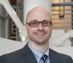
Task-Informed Evaluation and Design of Computational Imaging Systems
Mark Anastasio, PhD
Vice Chair of Imaging Sciences and AI Research
Director, Center for Computational and AI-Enabled Imaging Sciences (C2AIS)
Mallinckrodt Institute of Radiology
Associate Chief Research Information Officer for Biomedical Imaging, I2DB
Professor of Electrical and Systems Engineering
Washington University in St. Louis
Conventional image quality metrics such as peak signal-to-noise ratio (PSNR) and structural similarity index measure (SSIM) have long served as convenient surrogates for image reconstruction performance in computational imaging. However, these fidelity-based measures often correlate poorly with performance on the downstream tasks that ultimately matter, including detection, classification, and parameter estimation. I will discuss predictive v-information as a principled framework for evaluating and optimizing computational imaging systems according to the task-relevant information they preserve. From this perspective, imaging systems should be designed not merely to produce visually plausible reconstructions, but to maximize the information needed for accurate task performance.
I will also describe emerging approaches for incorporating task-based metrics directly into reconstruction method design, including training objectives and optimization strategies that explicitly align image formation with end-task success. Examples from inverse problems and learned image reconstruction will illustrate how this shift in perspective can reshape both the evaluation and design of computational imaging systems. Together, these ideas support a paradigm in which task performance, rather than visual similarity alone, defines image quality in computational imaging.
Nov. 2 - Jeff Nirschl, PhD | From Digital Pathology to PET Ligand Discovery: Computer Vision for Quantitative Tissue Imaging
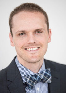
From Digital Pathology to PET Ligand Discovery: Computer Vision for Quantitative Tissue Imaging
Jeff Nirschl, MD, PhD
Assistant Professor, Department of Pathology
Neuropathology Core Leader, Wisconsin ADRC
PI, Wisconsin Brain Donor Program
University of Wisconsin-Madison
Neuropathology provides direct tissue-level evidence of disease, but extracting quantitative information from large collections of histology and autoradiography images remains difficult. Digital imaging and computer vision offer a way to transform these images into reproducible measurements while preserving their biological and spatial context.
This seminar will describe how we use computer vision across two complementary areas of neuropathologic imaging. First, I will discuss computational approaches for quantitative digital pathology, including segmentation, image registration, and spatial analysis of disease-associated tissue features. I will then describe how related methods are being applied to quantitative autoradiography for PET ligand discovery. In these experiments, brightfield histology and phosphor imaging are co-registered to measure radioligand binding within defined tissue compartments and to distinguish biologically meaningful signal from nonspecific binding.
Using examples from neurodegenerative disease, including efforts to develop ligands targeting aggregated proteins such as abnormal protein aggregates, we will show how pathology, image analysis, and molecular imaging can be combined to improve the scalability and interpretability of tissue-based screening. The broader goal is to use computer vision to augment expert interpretation, but as a quantitative bridge between microscopic pathology and in vivo imaging.
October 26 - David Dean, PhD | 3D Imaging, Virtual Surgical Planning, and Point-of-Care Manufacturing for Skeletal Reconstructive Surgery

3D Imaging, Virtual Surgical Planning, and Point-of-Care Manufacturing for Skeletal Reconstructive Surgery
David Dean, PhD
Professor of Biomedical Engineering, University of Wisconsin–Madison
In fall 2025, UW-Madison joined the NSF-funded HAMMER (Hybrid Autonomous Manufacturing, Manufacturing Evolution to Revolution) ERC (Engineering Research Center). With that move, the Center’s “Point-of-Care Manufacturing” projects were transferred to UW-Madison. HAMMER’s focus is to conduct mechanically informed Computer Aided Design of personalized devices. Once an optimized device is final, work shifts to engineering a fabrication process that will produce a device that meets those mechanical requirements. We will review novel Virtual Surgical Planning (VSP) software that allows this approach to be used to design personalized skeletal fixation devices. That VSP software offers the surgeon on a simulation of the anticipated post-operative biomechanical performance. In doing so, that biomechanical analysis, the software answers three critical questions the surgeon has: (1) what is the plate’s geometry, (2) what should it be made of, and (3) where should it be attached to the patient. In one example, those fixation plates are fabricated using robots at the point of care.
October 19 - Ariel Marshall, PhD | Beyond the Laboratory: Science, Policy, and the Expanding Role of the Scientist

Beyond the Laboratory: Science, Policy, and the Expanding Role of the Scientist
Ariel Marshall, PhD
Congressional Lead,
Scale AI
The American scientific enterprise was built through a partnership among the federal government, universities, national laboratories, and industry. The policies and institutions created under this model have shaped not only which research is supported, but also how generations of scientists have been trained and employed. Today, shifting federal priorities, evolving research institutions, and the growing role of technology across the economy are challenging traditional assumptions about what a scientific career looks like. This seminar will examine how public policy shapes the conduct and direction of science, explore the range of science policy careers available beyond the laboratory, and identify the transferable skills scientists need to help shape decisions across government, research institutions, and industry. Participants will leave with a broader understanding of how decisions about science are made and where their scientific training can position them to influence those decisions.
October 12 - Shannon O'Reilly, PhD & Jennifer Smilowitz, PhD | Global Health Initiatives and Opportunities in Oncology
Global Health Initiatives and Opportunities in Oncology
 Shannon O’Reilly, PhD, Associate Professor, Radiation Medicine, University of Wisconsin–Madison |
 Jeni Smilowitz, PhD, Clinical Professor, Radiation Medicine, University of Wisconsin–Madison |
Join us for an overview of the current global health initiatives in the Department of Radiation Medicine! We’ll highlight current programs and opportunities for you to get involved in global health. We will discuss building workforce capacity, advancing education, and strengthening access to radiation oncology care worldwide. We will present on some current experiences with the African School of Physics in Kenya, cervical cancer prevention education in Ghana and IMPACTO course in Colombia.
October 5 - Shawn Gomez, EngScD | Translational AI: Augmenting Clinical Decision-Making from Imaging to Therapy Design

Translational AI: Augmenting Clinical Decision-Making from Imaging to Therapy Design
Shawn M. Gomez, EngScD
Peter Tong Department Chair, Department of Biomedical Engineering
University of Wisconsin–Madison
Our group develops artificial intelligence and machine learning methods designed to augment, rather than replace, clinical decision-making across the continuum of care, from diagnosis to therapy selection. In medical imaging, we build deep learning models that extract clinically actionable information from routinely acquired images. For example, we have developed deep learning methods for intraoperative specimen mammography to predict tumor margin status during breast cancer surgery, with the goal of guiding resection and reducing repeat operations. On the therapeutic side, we combine large-scale drug perturbation data with tumor molecular profiles to predict how cancer cells respond to kinase inhibitors, alone and in combination. This helps prioritize the most promising treatments before they reach the lab or clinic. We also develop supporting methods for representing complex biomedical data and interpreting model predictions. Our unifying aim is to turn heterogeneous data, from images to molecular measurements, into trustworthy tools that are useful at the point of care.
September 28 - Chi Liu, PhD | Imaging Technology Developments in Quantitative PET and SPECT
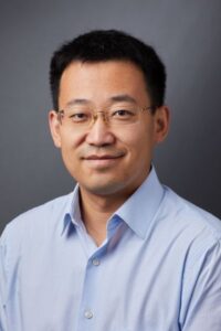
Imaging Technology Developments in Quantitative PET and SPECT
Chi Liu, PhD
Professor of Radiology and Biomedical Imaging, Associate Director of Yale Biomedical Imaging Institute
Yale University
This seminar will be covering topics of image reconstruction, motion correction, attenuation correction, and noise reduction for quantitative PET and SPECT imaging for the heart, cancer, and brain. AI-based imaging technologies will be focused.
September 21 - María-Ester Brandan, PhD | Quantification in Contrast-Enhanced-Mammography

Quantification in Contrast-Enhanced-Mammography
María-Ester Brandan, PhD
Professor, National Autonomous University of Mexico, UNAM
The speaker has coordinated the medical physics group at the UNAM Physics Institute for more than 20 years. One of the main projects has been the development of techniques and the accompanying formalism to quantify iodine uptake in radiological images of Contrast-Enhanced Mammography (CEM) using iodinated contrast media and dual-energy techniques. The seminar will present the project’s main achievements. We´ll discuss the methodology and structural adaptations required to implement pioneering medical physics investigations in a newly established clinical research environment. The participation of students has been essential, and we’ll include an overview of the UNAM master’s program on medical physics and its impact on strengthening the profession in Mexico.
September 14 - Sanhita Sinharay, PhD | Small-Molecule Zinc-Phthalocyanine Optoacoustic Agents -radiation-free metabolic imaging of peritoneal metastasis and a path to phototheranostics

Small-Molecule Zinc-Phthalocyanine Optoacoustic Agents -radiation-free metabolic imaging of peritoneal metastasis and a path to phototheranostics
Sanhita Sinharay, PhD
Assistant Professor, Indian Institute of Science
Detecting cancer where it is hardest to see, such as disseminated across the peritoneum or buried in deep-seated, treatment-resistant tissue, remains a central challenge for molecular imaging. The clinical mainstay for visualizing metabolically active tumors, 18F-FDG PET, delivers ionizing radiation, depends on costly radiopharmacy infrastructure, and resolves millimeter-scale disease poorly, and these constraints are especially limiting in resource-constrained settings.
This seminar presents small-molecule optoacoustic (photoacoustic) probes built on a zinc-phthalocyanine (ZnPc) scaffold as a versatile platform that aims to respond to that problem. I will focus on Glucose-Phthalocyanine (GPc), a first-of-its-kind, water-soluble, tetra-glucose-conjugated ZnPc that reports tumor glucose avidity, a radiation-free counterpart to FDG for imaging superficial tumors within a 5 cm depth. As a demanding test case, the presentation will focus on how GPc, imaged by multispectral optoacoustic tomography (MSOT), detects and longitudinally tracks peritoneal metastases in an intraperitoneal OVCAR-8 and OVCAR-3 ovarian cancer model, matched with the gross pathology and histology data.
I will then extend the platform from diagnosis to therapy: asymmetric ZnPc constructs that couple deep-tissue optoacoustic imaging with NIR-activated, ROS-generating photodynamic therapy, which we demonstrated in a pancreatic cancer model. Together, these results position the zinc-phthalocyanine scaffold as a single chemistry for translational imaging.