Medical Physics Seminar – Monday, October 6, 2014
Rapid and Accurate Single and Multicomponent Relaxometry Using Steady-State Acquisitions

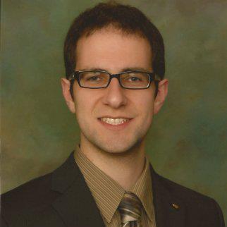
Samuel Anthony Hurley (student of Dr. Andrew Alexander)
Research Assistant, Dept of Medical Physics, UW-School of Medicine & Public Health, Madison, WI - USA -
Magnetic resonance imaging (MRI) is a medical imaging modality capable of generating images of the soft tissues of the human through the interaction of water molecules with their local tissue and microstructural environment. The signal observed in MR can be described by the parameters T1 and T2, and relaxometry is the study of making accurate and precise measurements of these fundamental parameters.
Unlike traditional methods such as inversion recovery and spin echo, which are slow and have limited spatial coverage, steady state sequences are an attractive choice for performing relaxometry experiments due to their short scan times and moderate to high-resolution volumetric coverage. In spite of these advantages, these methods suffer from a number of effects that reduce their accuracy, in particular strong dependence on the strength and variations of the radiofrequency (RF) excitation field. Two methods to measure and correct these RF effects, Actual Flip-angle Imaging (AFI) and Bloch-Siegert B1 mapping (BS), will be discussed.
Most relaxometry techniques measure a single value of T1 and T2 in each voxel. This is valid under the assumption of a single well-mixed pool of water, a condition that does not typically hold for the complex microstructural features of neural tissues. Multicomponent relaxometry is a technique to model multiple pools of water, each with its own T1 and T2 value. mcDESPOT is a method for performing multicomponent relaxometry, and will be discussed and demonstrated in vivo on an animal model of dysmyelination that exhibits an extreme paucity of myelin at birth.