Diffusion Tensor Imaging or DTI can be used in order to probe, in vivo, the intrinsic, three-dimensional diffusion properties of water within tissues. DTI has been applied in several studies to infer the microstructural characteristics of the brain (Pierpaoli et al., 1996; Virta et al., 1999), the heart (Holmes et al., 2000), and the spinal cord (Fenyes and Narayana, 1999). In addition, DTI has been introduced in the diagnosis of disease conditions such as cerebral ischemia (Lythgoe et al., 1997), acute stroke (van Gelderen et al., 1994) and multiple sclerosis (Werring et al., 1999). Unlike conventional DWI (Le Bihan, 1991), where diffusion-weighted (DW) images are used to calculate the scalar ADC, DTI characterizes diffusive transport of water by an effective diffusion tensor D. The eigenvalues of D are the three principal diffusivities and the eigenvectors define the local fiber tract direction field (Basser et al., 1994). Moreover, one can derive from D rotationally invariant scalar quantities that describe the intrinsic diffusion properties of the tissue. The most commonly used are the trace of the tensor (Basser et al., 1994, Pierpaoli et al., 1996, Basser, 1995), which measures mean diffusivity, Fractional Anisotropy (FA) and Lattice Index (LI)( Pierpaoli et al., 1996, Basser et al., 1995; Basser and Pierpaoli, 1996; Pierpaoli and Basser 1996), which characterize the anisotropy of the fiber structure, meaning how much higher the diffusivity is along some directions compared to others.
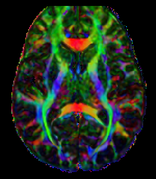
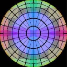
We use DTI in our research in an attempt to understand the underlying cause(s) of a variety of pathologies including epilepsy, traumatic brain injury, brain tumors and arterial-venous malformations. Shown below is an absolute value DTI colormap. In this visualization of the diffusion information, we assign colors to specific diffusion directions. X, Y and Z directions are assigned red, green, blue respectively. Any other direction within the xyz octant is assigned a combination of red, green and blue. Fibers with rotational or mirror symmetry appear to have the same color. Voxel brightness is scaled by its lattice index value. In order to determine the diffusion direction from this map, one uses the color wheel shown to the right. Assume the color wheel is actually a dome and you are standing at the center. The angle that your line of sight forms with a specific color on the dome represents the angular direction of the diffusion.
The diffusion pathways that are visible in the map are white matter fiber bundles. Diffusion in gray matter is more isotropic (no preferred diffusion direction) which is reflected in this map. DTI information can be obtained in 3D allowing fiber tracking throughout the brain.
References
- Pierpaoli C, Jezzard P, Basser PJ, Barnett A, Di Chiro G. Diffusion tensor MR imaging of the human brain. Radiology 1996;201:637-648.
- Virta A, Barnett A, Pierpaoli C. Visualizing and characterizing white matter fiber structure and architecture in the human pyramidal tract using diffusion tensor MRI. Magn Reson Imaging1999;17:1121-1133.
- Holmes AA, Scollan DF, Winslow RL. Direct histological validation of diffusion tensor MRI in formaldehyde-fixed myocardium. Magn Reson Med 2000;44:157-161.
- Fenyes DA, Narayana PA. In vivo diffusion tensor imaging of rat spinal cord with echo planar imaging. Magn Reson Med 1999;42:300-306.
- Lythgoe MF, Busza AL, Calamante F, Sotak CH, King MD, Bingham AC, Williams SR, Gadian DG. Effects of diffusion anisotropy on lesion delineation in a rat model of cerebral ischemia. Magn Reson Med 1997;38:662-668.
- van Gelderen P, de Vleeschouwer MHM, DesPres D, Pekar J, van Zijl PCM, Moonen CTW. Water diffusion and acute stroke. Magn Reson Med 1994;31:154-163.
- Werring DJ, Clark CA, Barker GJ, Thompson AJ, Miller DH. Diffusion tensor imaging of lesions and normal-appearing white matter in multiple sclerosis. Neurology 1999;52:1626-1632.
- Le Bihan D. Diffusion NMR imaging. Magn Reson Q 1991;7:1-30.
- Basser PJ, Mattiello J, Le Bihan D. MR diffusion tensor spectroscopy and imaging. Biophys J1994;66:259-267.
- Basser PJ. Inferring microstructural features and the physiological state of tissues from diffusion-weighted images. NMR in Biomedicine 1995;8:333-344.
- Basser PJ, Pierpaoli C. Microstructural and physiological features of tissues elucidated by quantitative-diffusion-tensor MRI. J Magn Reson B 1996;111:209-219.
- Pierpaoli C, Basser PJ. Toward a quantitative assessment of diffusion anisotropy. Magn Reson Med1996;36:893-906.